
Magnetic resonance imaging can be identified as a medical imaging technique. It can provide valuable details that are essential to diagnose some diseases, by visualizing certain soft tissue organs inside your body. Such as muscles, tendons, brain, nerves, and spinal cord can be visible. Before explaining further regarding how this test is performed, it is important to identify the technology behind this test.
The main two principles applied in this test are the establishment of a nuclear magnetic resonance and the application of radiofrequency pulses. Initially, the person will be placed in a magnetic field. Which will align the atoms in your body that have an electric charge such as water atoms. Then the radiofrequency pulses that are emitted will cause these atoms to emit signals which are detected and used to create images of the body. Sometimes a contrast agent is administered into your body. It will add before or during the procedure to enhance the visibility of certain structures or abnormalities inside your body.
Why is a MRI Done?
If you have any of the abnormalities mentioned below, your medical officer will recommend getting an MRI done.
MRI test will be useful to come for an accurate diagnosis and plan the management If you are having symptoms suspicious of below.
- A tumor in the brain or spinal cord
- Strokes
- Multiple sclerosis
- Parkinson’s disease
- Any lesion in cranial nerves
- this test will be useful to come for an accurate diagnosis and plan the management.
MRI will use to identify below mentioned disorders also.
- Muscles
- Bones
- Tendons
- Joints
- Ligaments or injuries to any of the structures
It is useful to diagnose and stage cancers in abdominal organs such as the liver, and kidneys, and pelvic organs (Bladder and prostate glands). Some gastrointestinal diseases and uterine conditions as endometriosis can also be diagnosed using MRI.
Most commonly MRI can be used to identify the exact location of any lesion in the spinal cord. Such as herniated discs, spinal cord compressions, spinal tumors, and any infections affecting the spinal cord.
If you are having,
- Urinary or fecal incontinence
- Pelvic organ prolapses
- Any other pelvic organ disorders
the strength of the muscle in the pelvic floor can be asses using MRI.
It is also the most appropriate imaging test used to identify below,
- Vascular abnormalities
- Malformations
- Any blood clots that get stuck in blood vessels.
Usage of this Test
- To diagnose the above conditions
- Plan the management
- Guide surgical interventions
- Find out the causes of those diseases
- Monitor the progression of those conditions
- Identify the response of those conditions to certain medications
How Should You Prepare for a MRI Scan?
- You need to follow a few important steps to make yourself ready to undergo this imaging technique to generate a successful imaging.
- You must follow all the instructions given to you by your medical officer.
- You must also inform your medical officer regarding any medical conditions or allergies you have and the surgeries you have undergone. Such as,
- Implanting metallic valves
- Pacemakers
- Aneurysm clips
Because these metallic parts inside your body can affect the images produced during an MRI scan.
- You normally don’t need to remain fasting before the MRI tests. But sometimes if you are undergoing an abdominal or pelvic MRI scan your medical officer will ask you to avoid taking food and drinking for a certain period before the procedure. Because of that, it is important to ask about the intake of food from your medical officer before the procedure and follow the instructions accordingly.
- You will also have to inform your medical officer if you are pregnant or breastfeeding. Although this is a safe procedure to be done during pregnancy it is important to inform your medical officer as they can take any special precautions considering your conditions.
- Your clothing can affect the quality of images produced. Hence it is important to wear comfortable loose clothing without any metal objects as zippers and buttons. Sometimes you will be advised to change into a hospital gown which is provided to you before the test.
- You will have to remove any jewelry, belts, watches, hair pins, eyeglasses, and such objects in your body that can interfere with the image quality before the test.
- There’s no reason for you to be afraid or anxious about the procedure. But if you feel that you are too stressed or anxious, you will have to inform your medical officer before the test as they can provide you with supportive methods such as sedation to support you to reduce anxiety and relax.
- Also, if it is a contrast-enhanced – MRI you will have some other special preparation steps to follow which are not followed in normal MRI imaging. You will have to remain fasting for a certain period and take some special medications to prepare yourself to receive a contrast agent.
What Happens During the Procedure?
- Initially your medical officer or the imaging technician will assess you for any metallic objects in your body and remove them.
- Next you will be offered a hospital gown to wear.
- Then you will be provided relaxation methods as sedation if you are anxious and in need of such methods.
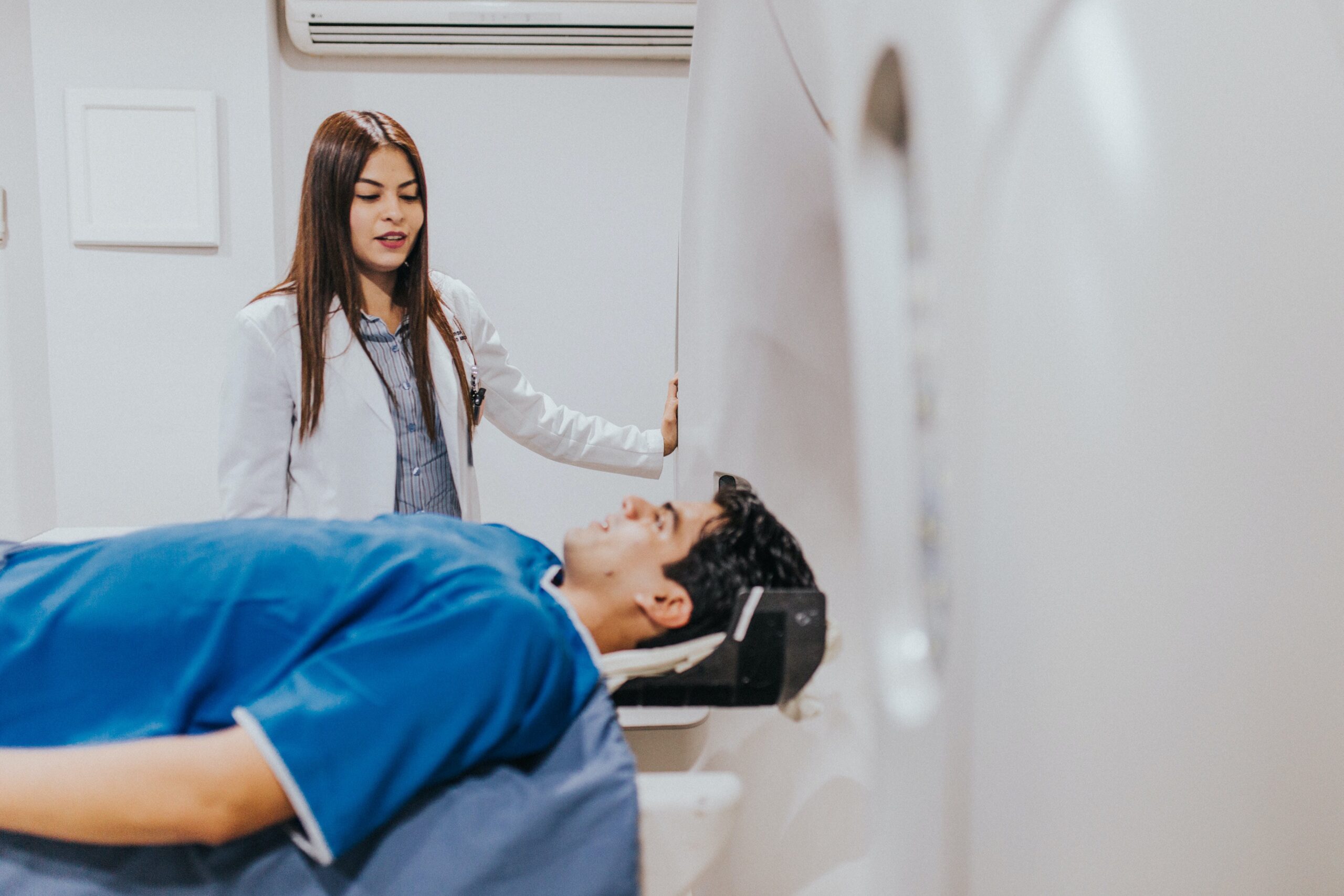
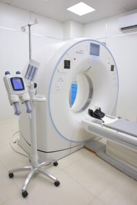
- You will have to lie on a table which moves into the MRI machine which has a bore where you will be placed during the procedure.
- Usually, you will have to lie in the face-down position and there will be an adequate cushioned surface in this table to support you to remain still and comfortable.
- Then the imaging will be performed and a loud buzzing sound of the machine will be heard throughout the procedure. You will be provided with earplugs or headphones with the facility to listen to your favorite music to distract yourself from hearing the noise of the machines.
- The technologist will communicate with you from the nearby control room by providing instructions and you will have the facility to inform the technician regarding any of your concerns to the technician during the procedure.
- The table you are lying on will slightly move during the procedure to get the best quality images.
Strictly Following Points
- The most significant responsibility you have during the procedure is remaining still and immobile as it is very important to generate a clear image.
- Normally the duration of a MRI scan can vary from 15 minutes to 1 hour duration.

What are the Limitations of MRI?
- The main limitation of MRI is the high cost and limited availability. Some patients are unable to afford such an expense while other imaging as X-ray and ultrasound scanning are relatively cheaper and widely available compared to MRI.
- This procedure takes longer than other procedures, making it difficult for patients to remain still and immobile for such long periods.
- Also, as the patients have to remain still inside the machine which is a confined space, they can experience anxiety and claustrophobia which is the fear of being in confined spaces. This requires the implementation of relaxation methods during the procedure.
- Some medical and psychiatric diseases make it difficult for some patients to undergo this scanning procedure.
- MRI scans also can’t get images of bones and air-filled spaces. Such areas in our body can interfere with the quality of the image produced by MRI.

References
- Kumar and Clerk’s Clinical Medicine -8th Edition- Parveen Kumar, Michael Clark
- Oxford Handbook of Clinical Medicine – 10th Edition
One thought on “Magnetic Resonance Imaging (MRI)”
Comments are closed.