

X-ray imaging is a common imaging investigation which had been used in the health sector for decades to view the inside of your body. X-ray imaging can be used to look inside your body without making any incisions. Which will help your medical officer to diagnose, manage and monitor diseases inside your body. Before further explanations about X-ray imaging, it is important to know what are X rays and how this imaging technique works.
X-rays are a form of electromagnetic radiation with powerful electromagnetic energy. Most of them have a wavelength ranging from 0.01 to 10 nanometers. It is corresponding to frequencies in the range 3 × 10¹⁹ Hz to 3×10¹⁶ Hz and energies in the range 100 eV to 100 keV. What happens in X-ray imaging is, that X-rays are produced when electrons coming in a high velocity collide with metal plates and produce energy in the form of X-rays. This energy which is produced in the form of X-rays gets absorbed by themselves into metal plates.
First X-ray beams are produced. After that it travel through the air and come in contact with the body tissues. Then it will produce an image on a metal film. The dense material inside your body as bones absorb X-ray beams. That is why their image can be produced on the metal film. But soft tissues like skin can’t absorb high energy. So rays as the x-ray beam passes through your body, hence images of such soft tissues can’t be produced in the image film.
Why is This Imaging Done?
- Your medical officer will recommend you get an X-ray imaging done when you are experiencing any of the following conditions.
- If you are experiencing pain or discomfort in an area of your body, diagnose the reason for the pain.
- If you have got any fractures in the back, upper limbs, or lower limbs identify the fracture and plan the management.
- To diagnose diseases that affect bones such as osteoporosis, and arthritis, and monitor the progression of such diseases. Monitor the response of such diseases to the drug treatments.
- To diagnose various conditions in the lungs such as pneumonia, effusions, lung abscess, etc.
- To identify breast tumors, cancer deposits in bones as well as bone cancers.
- To identify urinary or gallbladder stones.
Types of x-rays
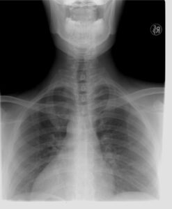
- Chest x-ray
- Abdominal x-ray
- Kidney, ureter, and bladder X-ray
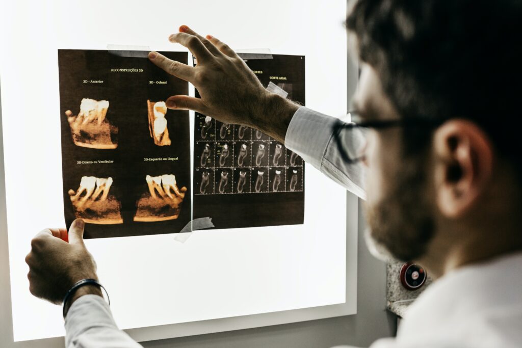
- Teeth and bone x rays
How Do You Prepare ?
- Usually, you don’t have to follow any special steps before the investigation.
- During the procedure you may be asked to change your clothes and wear a loose gown which is provided by the hospital.
- If not, you may be asked to wear loose comfortable clothing or adjust your clothing accordingly.
- Also, you will have to remove all the jewelry, belts, eyeglasses, watches, dentures, and any other metallic item you have in your body. As metallic buttons can also interfere with the imaging. So it is best to avoid wearing any clothes that include such material. You need to inform your doctor or radiologist if you are having any metal implants in your body. Such as metallic heart valves as these items can block the passage of X-rays through your body.
- You must also inform about any previous surgeries you have undergone or any medical conditions you have had during your life before the procedure.
- Normally X-ray imaging doesn’t require any special steps such as fasting. But if you are supposed to have x-ray imaging in the abdomen, your doctors may ask you to avoid eating any solid foods and restrict some liquid drinks as well for a certain period. Sometimes you will be prescribed medications that clean the bowel as well.
- It is also important to inform your medical officer if you are pregnant or currently breastfeeding. Because these rays can be harmful to the fetus and doctors might consider alternative methods to minimize the risk.
What Happens During x-ray Imaging?
- Normally the test is performed in a protected room and you will be asked to sit, lie or stand in several positions to get the proper imaging.
- If you are in the standing or sitting position you will have to stay in front of a specialized plate, that contains a sensor.
- You will have to stay in the asked position without moving for a few seconds to minutes. Remaining still as it is important to create clear images.
- If you are in a lying position, you may be asked to lie on a special plate and a camera will be moved connected to a steel arm over your body.
- After the procedure you can resume your daily activities and this procedure doesn’t need any hospital admissions.
- But if contract agents are used there will be certain steps you need to follow after the imaging and you may require hospital admission for a certain period with monitoring.
- Although this is a non-invasive and painless procedure. If you feel anxious or nervous during and after the procedure, you can follow steps. Engaging in deep breathing and any other relaxing techniques.
Limitations of x-ray Imaging?
- The main limitation is the use of ionizing radiation as it can have harmful effects on cells and tissues of your body. The risk that you can have from a single radiological imaging is very less. But if you get repetitive exposure to x-ray imaging you can have harmful effects and develop complications. This risk can be high for pregnant women and their babies. Hence special precautions need to be taken for them if they have to undergo this type of test.
- Unlike MRI scans and ultrasound scans, x-ray imaging can’t capture live or functional images. Hence, they provide fewer details regarding how organs are functioning. They only provide static images of structures inside of your body.
- They can’t capture images of soft tissues and only capture images of dense tissue as bones and teeth mainly. Also, they can’t capture images of the brain or spinal cord which has caused the need to do other tests such as MRI and CT scans.
- Also, X-ray imaging is not sensitive enough to capture early manifestations of diseases such as cancers and they can only be visualized in late stages.